IVD Gesellschaft für Innovative
Veterinärdiagnostik mbH

Parasitology

Parasitology deals with the interactions between parasites and their hosts, and in particular with the prevention, diagnosis and treatment of parasitic diseases. In addition to ectoparasites such as fleas, lice and mites, this also includes worm infections and infections caused by protozoa (single-celled organisms).

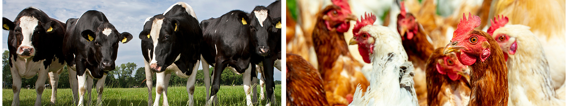
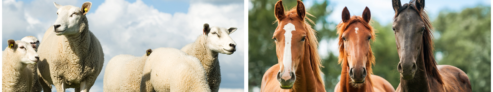
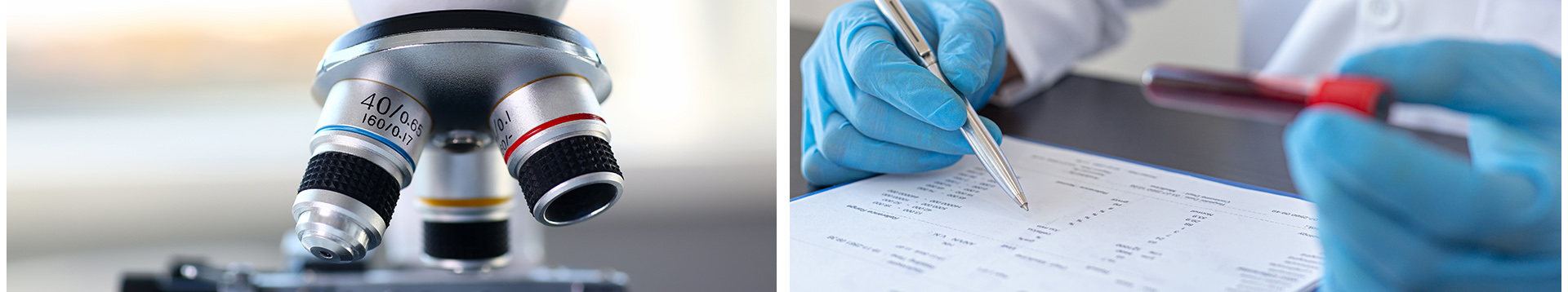
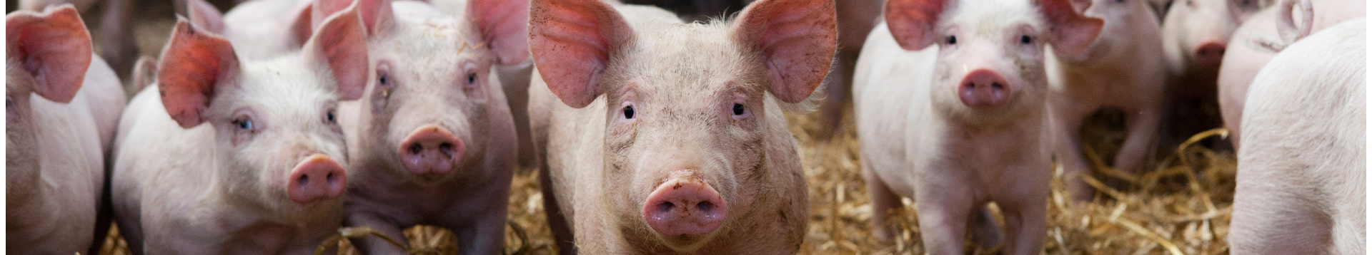
Since many parasites can cause serious illnesses in animals, but also in humans, efficient measures are required to combat parasites. The diagnosis essentially involves the microscopic assessment of the shape of the parasites or their eggs.

Microscopic Detection of parasite stages in faeces (Coproscopy):

Flotation

  • Concentration tequnique (enrichment) for the detection of eggs of cestodes (tapeworms) and nematodes  (roundworms, for example Ascaris suum in pigs, Cyathostominaein horses) as well as for the detection of protozoa (for example coccidia oocysts)
  • Material: collective fecal sample from 3 consecutive days due to possible intermittent excretion  (at least. 20-50 g)

 Sedimentation

  • Method for detecting heavy trematode eggs (flukes, for example , z.B. liver and rumen flukes in ruminants) as well as certain species of coccidia (for example Eimeria macusaniensis in New World camelids)
  • Material: collective fecal sample from 3 consecutive days due to possible intermittent excretion  (at least. 20-50 g)

 Combined sedimentation-flotation process

  • Higher sensitivity, particularly when detecting tapeworm eggs
  • More sensitive than the McMaster method due to the enrichment of the parasite stages before the detection step; thus detection of even low excretion of parasite stages

 Larval migration procedure (Baermann-Wetzel-method)

  • Is used to detect lungworm larvae
  • Material: collective fecal sample from 3 consecutive days due to possible intermittent excretion  (at least. 20-50 g)

Adhesive tape technique:

  • If infestation with Oxyuris equi (horse pinworm)  is suspected in horses
  • Samples from the horse's perianal region, as O. equi females deposit eggs in the perianal region and eggs can only be detected accidentally in the faecal examination
  • Microscope slide covered with a single layer of adhesive tape that has been applied to the horse's perianal area 

Faecal smear

  • Detection of cryptosporidia cysts in faeces (e. g. calf or lamb diarrheal) using carbol fuchsin staining
  • Material: fresh faecal sample (at least 5-20 g)

The material should be as fresh as possible and ideally stored cool (8-10 °C) to inhibit larval hatching and decomposition.

Examination for ectoparasites (e.g. lice, mites, fleas): 

Macroscopy and microscopy:

  • Depending on the location, deep skin scrapings, skin, hair, fur or feather samples or skin biopsies
  • Plumage sample on adhesive tape (3 pieces per bird in a sinlge layer on microscope slides)

Indirect detection by determining antibodies (e.g. ELISA for sarcoptic mange in pigs) as an expression of an infestation can also be helpful diagnostically.

Our diagnostic offers for various diseases and animal species can be found in our list of products and services as well as on the diagnostic request forms.